

You might think your eyes look healthy from the outside, but some of the most serious eye conditions develop silently in the back of your eye. Your retina, the light-sensitive tissue that sends visual signals to your brain, can show early warning signs of both eye diseases and serious health problems affecting your entire body. Lake Country Optometry uses retinal imaging technology to help detect these conditions early when treatment can be most effective.
Retinal imaging can detect diabetes complications, high blood pressure effects, macular degeneration, glaucoma, retinal detachment, and even brain tumors or stroke damage, often years before you notice any vision changes. Getting regular comprehensive eye exams with retinal imaging gives you the opportunity to catch these conditions before they affect your vision permanently.
How Retinal Imaging Works & What to Expect
Your Lake Country eye doctor uses digital cameras to capture detailed pictures of the back of your eye. The process takes about 5 to 10 minutes and feels comfortable, you simply position your face near the camera while it takes high-resolution images.
You’ll see a quick flash of light as the camera photographs your retina. In most cases, no dilation is needed, and the camera never touches your eyes. If your doctor needs an even closer look, they may recommend dilating drops for additional images.
These retinal photos help us detect early changes to your eye health with precision that a standard vision check can’t provide.
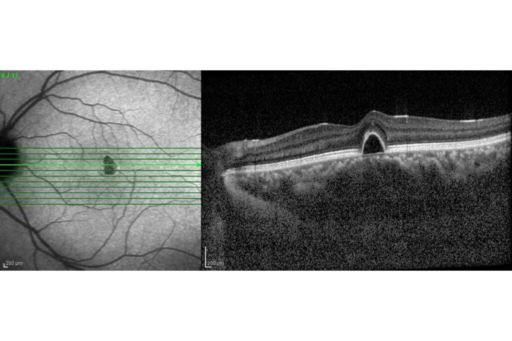
Types of Retinal Imaging
Your optometrist may use different imaging technologies depending on what they’re assessing:
- Retinal Photography: Captures wide-angle images of the retina, optic nerve, and blood vessels to detect visible changes.
- Optical Coherence Tomography (OCT): Uses light waves to take cross-section images of the retinal layers, helping diagnose conditions beneath the surface such as early macular degeneration or swelling.
Using both technologies together allows your eye doctor to detect even subtle signs of disease and compare changes from year to year.
Diabetes-Related Eye Conditions
High blood sugar damages the tiny blood vessels in your retina over time, leading to diabetic retinopathy. Your Lake Country optometrist can spot these changes through retinal imaging before you notice any vision problems. Regular diabetic eye exams help catch these changes early.
The images reveal several warning signs:
- Swelling or dilation of retinal blood vessels
- Blood or fluid leaking into your eye
- New abnormal blood vessels growing
- Fatty deposits under your retina
Understanding how diabetes affects your eyes can help you take the right steps to protect your vision through proper blood sugar management and regular eye care.
High Blood Pressure Effects on Your Eyes
Your retinal blood vessels show signs of hypertensive retinopathy when your blood pressure stays high for extended periods. These changes appear in retinal images before you experience vision problems or other symptoms.
Your eye doctor can identify these pressure-related changes:
- Narrowed or thickened blood vessels
- White cotton-wool spots indicating mini-strokes
- Fatty deposits on your retina
- Small hemorrhages in retinal tissue
Age-Related Vision Changes & Serious Eye Conditions
Macular degeneration affects your central vision and becomes more common after age 50. Your eye doctor at Lake Country Optometry can detect early signs through retinal imaging before your vision changes, giving you more treatment options. Early detection gives you more ways to slow progression and maintain your central vision.
Early detection markers include small protein deposits called drusen, retinal thinning in the macula area, fluid buildup under your retina, and changes in retinal pigmentation.
Glaucoma also shows up in retinal images through changes in your optic nerve appearance and pressure effects on nerve fibers. Your eye doctor can track progression by comparing your images year to year. Learn about the symptoms and treatment options for glaucoma to better understand this silent vision thief.
Signs of Serious Health Problems Beyond Your Eyes
Your retinal images can reveal health conditions affecting your entire body. The blood vessels in your eyes often show the first signs of systemic diseases, making retinal imaging a valuable health screening tool.
Brain and nervous system issues appear through swollen optic nerves from increased brain pressure, signs pointing to potential tumors, optic nerve damage from multiple sclerosis, and effects of stroke on your visual system. Some conditions detected through retinal imaging need prompt treatment to prevent permanent vision loss or other serious complications.
Protect Your Vision With Early Detection
Retinal imaging gives your Lake Country Optometry team a detailed record of your eye health. These images create a baseline we can compare year after year, helping us spot even the smallest changes before they affect your daily life. When conditions are detected early, treatment is often simpler, less invasive, and far more effective. Regular imaging is a smart investment in your long-term vision.
Whether you are managing diabetes, have a family history of eye disease, or simply want peace of mind, advanced retinal imaging is one of the most powerful tools we have to protect both your sight and your overall health.
Book your comprehensive eye exam in Lake Country today and see how modern diagnostic technology can help keep your eyes healthy for years to come.